Extinct Tasmanian Tiger now back in 3D
Using 3D scanning, researchers are peeking under the preserved skin of Tasmanian tiger specimens to reconstruct its growth and development.

When all that remains of an extinct species is a few precious specimens, dissecting them, even in the name of science, isn’t really an option.
But by using CT scanning, a technique similar to medical CAT scans but with much higher resolution, researchers from the University of Melbourne and Museums Victoria have been able to take a ‘virtual’ look inside the Tasmanian tiger to study the biology of this iconic species.
Called non-invasive X-ray micro-CT scanning, this technique has also been used to examine Egyptian Mummys, another example of rare and delicate specimens.
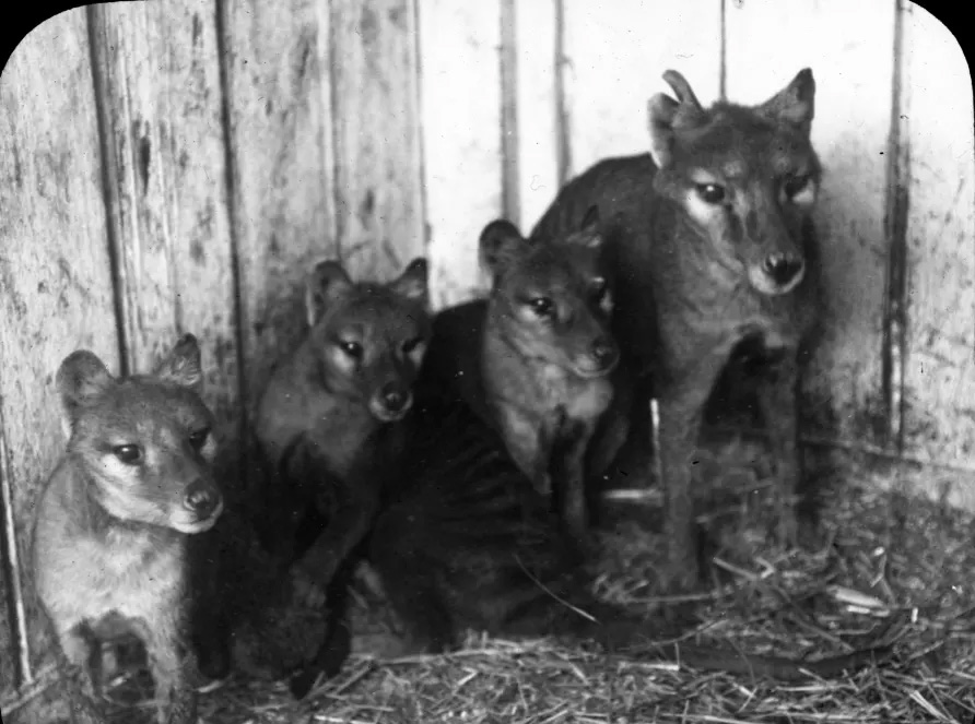
Dr Christy Hipsley, Research Associate at Museums Victoria and the University of Melbourne says that before they were hunted to extinction in 1936, it was very popular for museums to collect samples of the Tasmanian tiger, also known as thylacine or Thylacinus cynocephalus.
Limited Specimens Preserved
She says that a very limited number of pouch young specimens (joeys) were collected and preserved, and these now exist in various museum collections across the world from Tasmania to Prague.
“Due to the technological limitations at the time the thylacine went extinct, there are only limited details on its growth and development,” Dr Hipsley says.
The research team conducted CT scanning on all 13 known pouch young world-wide to create 3D digital models. The models have enabled the team to study their skeletons and internal organs, and reconstruct their growth and development.
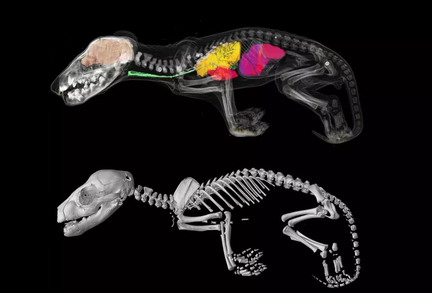
Axel Newton, a University of Melbourne PhD student and lead author on the paper, notes that the collection of joey specimens represents five critical stages of postnatal, or pouch development.
“Our 3D models have revealed important new information about how this unique extinct marsupial evolved to look so similar to dogs, such as the dingo, despite being very distantly related,” says Mr Newton.
From Joey to puppy
“The digital scans show that when first born the Tasmanian tiger looked like other marsupials like the Tasmanian Devil or the kangaroo.”
These scans show in incredible detail how the Tasmanian tiger started its journey in life as a joey boasting the robust forearms of other marsupials so that it could climb into its mother’s pouch. But by the time it left the pouch around 12 weeks to start independent life, it looked more like a puppy, with longer hindlimbs than forelimbs.
The Tasmanian tiger’s resemblance to the dingo is known as one of the best examples of convergent evolution in mammals. This is where, two species, despite not being closely related, evolve to look very similar. The Tasmanian tiger would have last shared a common ancestor with the canids (dogs and wolves) around 160 million years ago.
Dr Christy Hipsley says after sequencing the Tasmanian tiger genome in 2017, this research is one more piece in the puzzle of why they evolved to look so similar to dogs.
Once ranging throughout Australian and New Guinea, the Tasmanian tiger disappeared from the mainland around 3,000 years ago, likely because of competition with humans and dingos.
The remaining Tasmanian tiger population, isolated on Tasmania, was hunted to extinction in the early 20th century, with the last known individual dying at Hobart Zoo in 1936.
Associate Professor Andrew Pask from the University of Melbourne explains the scanning was an incredibly effective technique to study the skeletal anatomy of the specimens without causing any damage to them.
“This research clearly demonstrates the power of CT technology. It has allowed us to scan all the known Thylacine joey specimens in the world, and study their internal structures in high resolution without having to dissect or cause damage to the specimen,” Associate Professor Pask says.
Dissection not an option
“By examining their bone development, we’ve been able to illustrate how the Tasmanian tiger matured, and identify when they took on the appearance of a dog.”
The study has also revealed that two specimens held in the collection of the Tasmanian Museums and Art Gallery (TMAG) weren’t Tasmanian tigers at all. Instead, they are most likely to be quolls or Tasmanian devils, based on the number of vertebrae and presence of large epipubic bones (the specialised bones that support the pouch in modern marsupials).
Senior Curator of Vertebrate Zoology at TMAG, Ms Kathryn Medlock says the museum has received many requests to dissect its pouch young over the years but they are always refused.
“One of the major advantages of this new technology is that it has enabled us to do research and answer many questions without destruction of the sample specimens,” Ms Medlock says.
“This is a significant advancement that also has an additional benefit of helping us to learn more about the identity of these specimens that have been in the TMAG collection for many years.”
The 3D digital Tasmanian tiger models are publicly available here as a resource for current and future researchers.
Written By Dr Nerissa Hannink, University of Melbourne
This article was first published on Pursuit. Read the original article.

80‑million‑year‑old snake fossil sheds light on why lizards lost their limbs and started slithering
Snakes are everywhere in our legends and mythology. Yet for most of us, our blood runs cold whenever we encounter these strangely undulating, scaly tubes of muscle slithering through the leaf litter.

An extinct echidna the size of a small child once roamed Victoria, new fossil shows
More than 100 years after it was found in Foul Air Cave in Victoria, the fossil is grating us new insights into deep time.
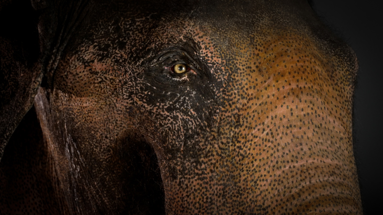
The Journey of Bong Su
How taxidermy honours a beloved Asian Elephant